The first published scientific study of Neurospora, including a description of photoinduction
of carotenoids
D.D. Perkins - Department of Biological Sciences, Stanford University, Stanford, CA 94305-
5020
In the warm, moist summer of 1842, bread from army bakeries in Paris was spoiled
by massive growth of an orange mold. A commission was set up by the minister of war to
investigate the cause of the infestation and to make recommendations. Their report (Payen
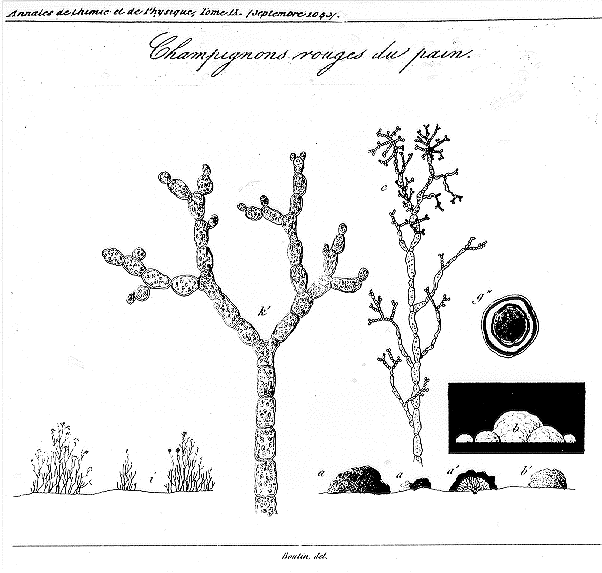
1843) includes a colored plate which shows mycelia, conidia and colonies of the
"Champignons rouges du pain" (called Oidium aurantiacum). I have translated one passage
which concerns the effects of illumination:
"With the object of determining if the coloration was due to light, even extremely
dim, we attempted to exclude light completely by putting a piece of bread in a glass
flask containing 10 grams of water. The flask was surrounded by black paper and
enclosed in a vessel of half-centimeter thick bronze.
Development of the fungus was a little less abundant than on a piece of the
same bread that was exposed to light under conditions that were otherwise identical.
Under the first conditions, the fungi remained completely white for more than eight
days (see figure b), whereas the illuminated fungi, figure a, a', were covered with red
spores. But, remarkably, the white fungi became colored when they were exposed to
light for two hours."
The 1843 report names Lévillé, Montagne and Decaisne as scientists concerned with identifying
the organism, while de Mirbel and one of the scientific members of the commission (Dumas, Pelouze
or Payen) were concerned with microscopic and chemical analysis. Montagne (1843) independently
published a drawing of the same orange fungus with a Latin description under the name Penicillium
sitophilum.
Thermal tolerance of Neurospora was also studied at about this time (see Payen 1858, 1859).
The orange spores survived 100 C for one hour and exposure to 120 C for an unspecified period. They
were killed, however, at 140 C. These results were cited by Louis Pasteur (1861) in a paper reporting
that mold spores could survive dry heat at 120 C to 125 C for at least an hour whereas viability was lost
in a few minutes if the spores were heated in water at 100 C. These observations were relevant to the
controversy then current regarding spontaneous generation.
Montagne, C. 1843. Quatrième centurie de plantes cellulaires exotiques nouvelles. Ann. Sci. Nat.
Bot. 2e Sér. 20:352-379 (+ one plate).
Payen, A. (rapporteur) 1843. Extrain d'un rapport addressé à M. Le Maréchal Duc de Dalmatie,
Ministre de la Guerre, Président du Conseil, sur une altération extraordinaire du pain du
munition. Ann. Chim. Phys. 3e Sér. 9:5-21 (+ one plate).
Payen, A. 1848. Températures qui peuvent supporter les sproules de l'Oidium aurantiacum sans
pedre leur faculté végétative. Compt. Rend. Acad. Sci. 27:4-5.
Payen, A. 1859. [Untitled discussion following remarks of M. Milne Edwards rejecting spontaneous
generation of animalcules.] Compt. Rend. Acad. Sci. 48:29-30.
Pasteur, L. 1862. L'influence de la temp‚rature sur la fécundité des spores de Mucédinées. Compt.
Rend. Acad. Sci. 52:16-19.

Excerpt from Plate 1 of Payen (1843). The original plate is in color. a. Colonies of the red-
orange fungus Oidium aurantiacum as they appear to the naked eye in the cavities of infected bread.
a'. A similar colony cut in two, showing in the red area a thick layer composed of innumerable small
spores formed at the end of radiating filaments. The latter are yellowish white. b. Similar colonies
that have grown up completely in the dark, with the result that the red color has not developed. b'.
One of the colonies in b seen after exposure to light for one hour. Color begins to appear and then
pigmentation progresses rapidly. c. Branching filament, about 150 x. g". Spore treated successively,
under the microscope, with a dilute solution of potassium hydroxide, and aqueous alcoholic solution
of iodine, then with gradually more concentrated solutions of sulfuric acis. This acid, which separates
parts of the cellulose envelope that contains less nitrogenous substance, results in a blue color turning
to purple, which is characteristic of the state intermediate between cellulose and dextrin. i. Normal
vegetative growth as seen with the naked eye; well developed, especially under conditions of high
humidity. k'. Termini of well developed filaments, showing spores and young cells.
