
Click picture for larger image
a) the hyphal "cell"
b) the conidium
c) the ascospore
All Neurospora cells contain the basic components of a eukaryotic cell: nuclei, mitochondria, endoplasmic reticulum, golgi, vacuoles, various types of vesicles, cytoskeleton, ribosomes and others. The cytoplasm is enclosed by a plasma membrane and outside this is a rigid cell wall largely composed of the polysaccharide chitin.
At least a dozen specialized cell types can be distinguished at different stages of the life cycles, but three cell types are the most prominent:
Click picture for larger image
a) the hyphal "cell"
b) the conidium
c) the ascospore
Hyphae
Hyphae are tubes with walls that are rigid except at the tips, which is where growth occurs. Cell wall and plasma membrane components are synthesized some distance from the tip and transported forward to the tip in vesicles, along the strands of the cytoskeletal "highway". The vesicles accumulate at the tip in a body called the Spitzenk÷rper, which seems to distribute the vesicles to the apical region of extension. A tip-high gradient of calcium ions is needed for growth to occur. New tips emerge as lateral branches some distance behind the tip.
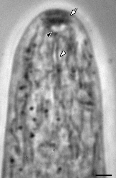
Neurospora crassa
wild-type hyphae shown with phase contrast optics.
A well-organized Spitzenk÷rper
composed of a
core (black arrowhead),
secretory vesicles (white arrow) and functional protein (not shown).
Mitochondria (white arrowhead) are visible.
Scale bar ~ 1 mm.
(Image by M. Uchida and R.W. Roberson)
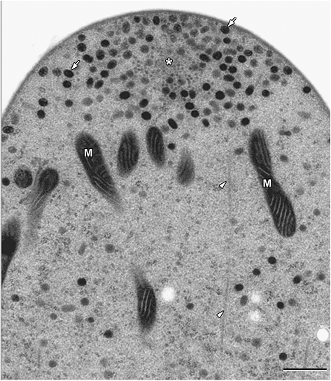
Transmission electron microscopy of cryofixed and freeze-substituted wild-type
hyphae of Neurospora crassa. Near-median longitudinal section
through hypha
illustrating the Spitzenk÷rper was composed of a cloud of secretory vesicles
(arrows)
surrounding a core (asterisk). Microtubule (arrowheads) and mitochondria (M)
are noted.
Bar = 3.3 mm. (Image by R.W. Roberson
from: Riquelme, M., Roberson, R.W. McDaniel, D.P.,
Bartnicki-GarcĒa, S., 2002. The effect of ropy-1 mutation on cytoplasmic
organization in
mature hyphae of Neurospora crassa. Fungal Genetics and Biology 37:
171-179)

GFP tagged hypha of N. crassa
Courtesy of N. L. Glass
Conidia
The conidia most often dealt with (because they are most prominent) are macroconidia. These are spheroid cells, with an average of 2 to 3 identical nuclei, that can live up to several weeks at room temperature (longer when refrigerated). They are units of dispersal. Under the appropriate conditions a conidium initiates polar growth by sending out a germ tube which then becomes the first hypha.
Conidia also act as paternal fertilizing elements (spermatia) in heterothallic species. When applied to the opposite mating type, a sex hormone emitted from the conidium attracts a fertile hair (trichogyne) from the maternal parent, and the two fuse as a prelude to the sexual cycle within the perithecium.
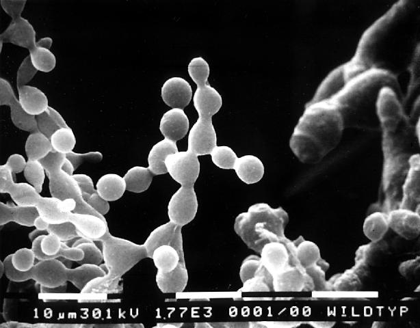
SEM micrograph of conidia of N. crassa
photo courtesy of M. Springer
Ascospores
These football (rugby ball) -shaped cells with thick black walls develop from the products of meiosis. Each has numerous identical haploid nuclei. Ascospores are also units of dispersal, but differ from conidia in being more resistant to environmental stress (such as desiccation), and in being heavier hence traveling shorter distances in air. Heat, or chemicals produced by heating the substrate, will induce germination. Polarized growth is initiated and a germination tube (hypha) emerges.
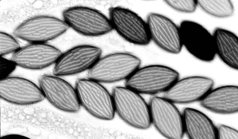
ascospores of N. crassa
Photo courtesy of N. Raju
Last modified 4/28/04 KMC