Presence of double stranded RNA in natural isolates of Pycnoporus cinnabarinus
Cifuentes, V., C. Martinez and G. Pincheira Laboratorio de Genetica, Dpto. de Ciencias Ecologicas,
Facultad de Ciencias, Universidad de Chile, Casilla 653, Santiago, Chile.
While studying the nucleic acids of different strains of P. cinnabarinus (Pc58, Pc470.3, Pc470.6), the presence of dsRNA molecules (1.7 kb to 3.6 kb) was detected in two of them.
Pycnoporus cinnabarinus is a basidiomycete capable of degrading lignocellulose and is known as a
white rot fungus. P. cinnabarinus strains Pc58, Pc470.3 and Pc470.6 were isolated from carpophores
collected in the rain forest of southern Chile. In the laboratory these strains were grown in liquid PC
medium for 15 days at 30°C without shaking. The mycelia were harvested on Whatman #1 paper and
washed with distilled water. Then the mycelia were frozen at -20°C and ground in a mortar. Nucleic acids
were prepared by the rapid lithium chloride method (Leach et al. 1987 FGN 34:32-33), except that three
phenol:chloroform:isoamyl alcohol (25:24:1) and two chloroform:isoamyl alcohol (24:1) extractions were
performed before precipitating with ethanol. Nucleic acids were resuspended in sterile distilled water and
later were dialyzed against TE buffer (10 mM Tris-HCl, 1 mM EDTA) for 16 h. The preparations were
than analyzed by electrophoresis on a 0.7% agarose gel followed by staining with ethidium bromide.
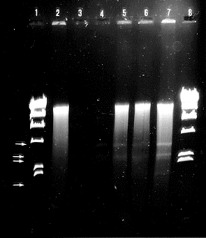
The electrophoretic analysis showed that strain Pc58 has
only chromosomal DNA. Strains Pc470.3 and Pc470.6 have
four bands moving faster than chromosomal DNA (Fig. 1).
These bands correspond to dsRNA as determined by
different tests with nucleases. These dsRNAs, denominated
as L-dsRNA, M1-dsRNA, M2-dsRNA and S-dsRNA, have
been purified by electrophoresis in low-melting-point
agarose followed by CTAB extraction (Langridge et al. 1980
Anal. Biochem. 103:264-271). Their molecular sizes were
approximately 3.6 kb, 2.7 kb, 2.3 kb and 1.7 kb respectively.
P. cinnabarinus strains Pc470.3 and Pc470.6 seem to
have the same types of dsRNA. Furthermore, the presence
of different dsRNA in the same strain has been observed as
in other fungi (Myers et al. 1988 Curr. Genet. 13:495-501).
Since dsRNA is an indicator of the presence of viruses
in fungi, our observations in P. cinnabarinus suggest the
presence of a virus-like particle which does not seem to be
associated with a visible alteration in phenotype of the
fungus.

Figure 1. Agarose gel electrophoresis of nucleic acid
preparations from natural isolates of P. cinnabarinus. Lanes
1 and 8: DNA digested with endonuclease HindIII; lane 2 strain Pc58; lane 4 strain Pc470.6 digested
with DNAse I; lanes 5 and 6 strain Pc470.3; Lane 7 strain Pc470.6
Return to the FGN 37 index
Go to the FGSC main page
