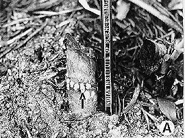
Fig. 1.A. Conidiating pustules of Neurospora (arrow) emerging through root openings at the node of a scorched sugar cane stump (approximately two weeks after the burn).
Except for a solitary report on the presence of perithecia of Neurospora on pine trees burned in the fire which followed an earthquake in Tokyo (Kitazima 1925 Ann. Phytopath. Soc. Japan 1:15-19), all other attempts to find the sexual stage in nature were unsuccessful (Shear and Dodge 1927 J. Agric. Res. 34:1019-1024; Prakash 1965 Neurospora Newsl. 7:5-6; Shaw 1990 The Mycologist 4:6-13). However, based on the available information on the population structure of Neurospora, Perkins and Turner (1988 Exp. Mycol. 12:91-131) predicted that sexual reproduction is prevalent in nature.
We present evidence for the occurrence of sexual reproduction of Neurospora in nature. Because of the preference of Neurospora for burned substrates and the regular availability of scorched sugar cane in agricultural fields, this material was selected for examinations. The place of our study was Maddur in Mandya District, approximately 80 km southwest of Bangalore. After sugar cane is reaped, the trash of the stripped leaves left on the ground is burned to clear the fields. The sugar cane stumps are scorched, but the development of new shoots (ratoon crop) from the underground portions of these stumps is not affected. The calendar of events in sugar cane fields which had been burned in January was as follows:
About a week after the fields were burned, Neurospora was visible as distinct pink conidiating pustules which were more common at the nodes than at either internodes or the cut surfaces of the scorched stumps (Fig. 1A). The mycelium ramified in the stump and was dense under the epidermal plant tissue. Pustules of conidia emerged at the nodes through root openings and through cracks in the epidermal tissue. The juicy stumps supported profuse and prolonged conidiation. These stumps were brought to the laboratory and examined with a binocular microscope. Perithecia of Neurospora were not found in the stumps in which Neurospora was conidiating.
One to two months after the burn (February-March), the stumps had begun to shrivel. These were surrounded by a ratoon crop which had grown approximately two feet high. Pale and withered pustules of Neurospora could still be seen on the stumps. However, these stumps also did not have perithecia either on the surface or under the epidermal tissue.
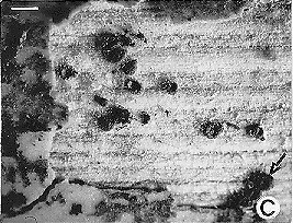
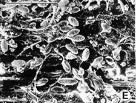
By the end of April, the new crop had grown approximately three feet, obscuring the stumps. Although pre-monsoon showers had come in the last week of April, the burned- over stumps appeared dry. In these shrivelled stumps, the epidermal plant tissue had become separated from the ground tissue and had begun to fall off (Fig. 1B). Excepting old, whitish patches of Neurospora in a few stumps, the conidial pustules had mostly disappeared. When epidermal tissue was peeled off, clusters of Neurospora perithecia were found at a few places on the ground tissue in some stumps (Fig. 1C). Most perithecia were shrivelled but a few were typically flask-shaped with beaks (Fig. 1D). In some stumps perithecia protruded through the cracked epidermal tissue. Some perithecia contained asci in different stages of development. In a few stumps, perithecia had ejected ascospores, which were sticking to the under surface of the epidermal tissue or on the ground tissue (Fig. 1E). The ascospores were grooved and measured 27 x 15 m. When ascospores were isolated on Vogel's N medium + 1.5% glucose and heat-shocked, nearly 40% germinated. The resulting cultures were identified as Neurospora intermedia, based on successful crosses with tester strains.
We have observed perithecia in burned-over stumps of sugar cane for three successive years. The observation that asexual and sexual stages of Neurospora are temporally separated and that perithecia are found under the epidermal tissue has raised questions as to the role of conidia, the mechanism of fertilization, the mode of dispersal of ascospore and the mechanism of initiation of 'infection'. Information on these and other aspects of the life cycle of Neurospora in nature will be published elsewhere.

Fig. 1.A. Conidiating pustules of Neurospora (arrow) emerging through root openings at the node of a
scorched sugar cane stump (approximately two weeks after the burn).

Fig. 1B. Shrivelled stump (arrow) surrounded by a ratoon crop approximately three months after the burn.

Fig. 1C. Light micrograph of perithecia on ground tissue partially exposed by removing epidermal tissue of the burned sugar cane stump. Perithecia (arrow) were those of Neurospora. Other perithecia were of Gelasinospora as identified by ovoid ascospores with pits under SEM. (Bar, 1 mm).

Fig. 1D. SEM of Neurospora perithecia (arrow) on ground tissue of burned sugar cane stump. (Bar, 1 mm).

Fig. 1E. SEM of Neurospora ascospores on a stump. (Bar, 100 microns).
Acknowledgements. We thank David Perkins, Stanford University, for inspiration. This work was supported by a grant from Department of Science and Technology, Government of India.