A non-radioactive electrophoretic mobility shift assay for the detection of heat shock element (HSE)-binding activity in Neurospora crassa - U. Meyer and L. Rensing Institute of Cell Biology, Biochemistry and Biotechnology, University of Bremen, NW2, Leobener Str., 28334 Bremen, Germany.
A non-radioactive electrophoretic mobility shift assay based on the digoxigenin detection system had been developed and applied to the analysis of the interaction between the heat shock transcription factor and the heat shock element of N. crassa.
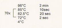
The labeling reaction was done with a thermostable Taq-polymerase (Biotherm, Genecraft, Germany) in a thermocycler under the following conditions. 1)10 x polymerase-buffer 2l, 2) stock solution of dATP(2mM), dCTP(1mM), dGTP(1mM) 4 l, 3) digoxigenin-dUTP(1mM) 1 l, 4) HSE (0.5 g/l) 1 l, 5) H2O 11.5 l, 6) polymerase (5U/l) 0.5 l.
Protocol for the thermocycler : A hotstart is recommended; add 50 l mineral oil to cover the sample.
In this protocol the annealing temperatures are higher than the elongation temperature (72C). Choosing the annealing temperature lower than 72C would lead to a DNA-probe, which is not HSE-like. During this labeling-reaction dTTP is substituted by digoxigenized dUTP (Boehringer Mannheim, Germany). It is important to use a polymerase that accepts dUTP as a dTTP analog. Following the label reaction, the mineral oil was discarded, and the probe was precipitated by adding ethanol (80 l) in the presence of LiCl (2.5 l of a 4 M solution) and stored overnight at -20C. The probe was then centrifuged at 18,000 g (4C) for 30 minutes. The supernatant was discarded and the precipitate resolubilized in 50 l bidistilled water. Serial dilutions were dot-blotted onto a DNA-binding membrane to check the label-efficiency. The probe should be detectable even at amounts as low as 0.2 fmol. Our probe was usually detectable at even lower amounts (0.02 fmol).The probe was stored in aliquots at -20C. We used 1l (200fmol HSE) of the above mentioned solution for one EMSA sample preparation.
N. crassa (bd, mat A strain, FGSC-stock no.1858) homogenate was prepared according to the modified procedure for the S. cerevisiae heat shock transcription factor (Sorger and Pelham 1987 EMBO J. 6:3035-3041). N. crassa mycelium was harvested from liquid cultures (9 cm petri dish, 40 h after inoculation with 1.3x10⁶ conidia/25ml), the medium was then removed, and the mycelium quickly frozen in liquid nitrogen. The mycelium was ground in a precooled mortar with sea sand for about 2-3 minutes. The resulting powder was placed into a small glass vessel, and 1 ml of homogenization buffer (200 mM Tris-HCl pH 8.0, 10% glycerol, 10 mM EDTA, 10 mM DTT) was added.
The mixture was stirred on a magnetic stirrer for 20 minutes at 4C and the homogenate centrifuged for 30 minutes at 18,000 g. The supernatant was removed and recentrifuged at the same conditions and the resulting supernatant used for the gel shift assay after determining the protein concentration (25 g of whole cell extract was used to perform one assay). The homogenate was stored at -20C. 5 x Binding buffer was prepared according to Sorger and Pelham 1987 EMBO J. 6:3035-3041, modified; 1x : 20 mM HEPES-KOH pH 7.9, 60 mM KCl, 12 % glycerol, 2 mM EDTA, 2 mM DTT. The nonspecific competitor consisted of poly d(I-C) 0.5g/l or plasmid DNA, e.g. pBR322, the specific competitor of 400 ng/l unlabeled HSE-probe (Figure1b).
The solutions were mixed in the described way (Table 1), vortexed and centrifuged briefly. The binding reactions were incubated for 20 min at room temperature and the samples loaded onto a 4% native 1x TAE-polyacrylamide gel. The samples were electrophoresed at 200 Volts for about 1.5 h in a cold room and the gel was electroblotted onto a membrane (a positively charged nylon membrane is strongly recommended, e.g. Amersham Hybond-N+) at 400 mA for 60 min. This is followed by UV-crosslinking and/or baking (80-100C for 30 min). The digoxigenized probe was detected by an anti-digoxigenin-fab-fragment conjugated with alkaline-phosphatase according to the manual of Boehringer Mannheim. We always used the BCIP/NBT dye system to detect signals.
The results showed that the non-radioactive EMSA is suitable to examine HSE-specific band shifts in N. crassa. The experiments revealed a gel shift pattern similar to that of S. cerevisiae, showing a specific constitutive HSE-binding activity under normal growth conditions (Figure 2: Lane 1). This signal decreased in heat shocked mycelia (41C for 1 h). Instead, a more retarded specific signal appeared (possibly due to HSF-phosphorylation during the heat shock) (Figure 2: Lane 2), which is again similar to S. cerevisiae. A heat shock of 43C totally erases the constitutive signal but leads to an increased signal strength of the above mentioned slower migrating band (data not shown). We assume that these signals represent interactions between the HSE-probe and the HSF of N. crassa.
Table 1: Scheme of binding reactionsa.
| Lanes | 1 | 2 | 3 | 4 |
| dig.-HSE-probe | + | + | + | + |
| poly d(I-C) | + | + | + | + |
| specific competitor | - | - | + | + |
a The 5 x binding buffer (3l) is first pipetted into a reaction tube, followed by distilled water (ad 15 l), poly d(IC) (1 l; 500 ng) and the digoxigenized-HSE-probe (1 l; 200 fmol). The specific competitor (1 l; 400ng) was used in binding reaction 3 and 4. Finally 25 g (different volume) of whole cell extract was added.
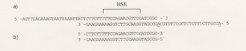
Figure 1: a) HSE-probe from S. cerevisiae. The nucleotide sequence within the bracket is the HSE itself. Instead of C and T, two adenosines (both are underlined) were inserted into the sticky end on the right, to enable the incorporation of digoxigenin-dUTP. b) Unlabeled HSE was used as a specific competitor to distinguish between specific and nonspecific signals.

Figure 2: Non-radioactive electrophoretic mobility shift assay with cell extracts from N. crassa.
Lanes 1+3 = cell extract from control cells; lanes 2+4 = cell extract from heat shocked cells; fp = free probe; c = constitutive HSE-binding activity in control mycelia; hs = HSE-binding activity in heat shocked mycelia. ns = non-specific signal; HSE specific signals are no longer detectable in lanes 3+4 due to the presence of specific competitor.